The lower urinary tract (LUT) system consists of the bladder and urethra and dysfunction of lower urinary tract leads to urinary incontinence, voiding dysfunction or both. The storage and emptying of bladder being a fundamental physiological activity, diagnosis of storage and emptying disorders of bladder is usually clinical. (1)
The lower urinary tract symptoms are very closely related to the bladder pressure. The fluid naturally flows from a point of high pressure to a point of low pressure. If high pressure sets up in bladder, urine leak may occur and at times of low pressure in bladder, there will be difficulty in bladder emptying. Urodynamic study is the dynamic invasive pressure study to assess the various parameters of the LUT to assess its function and dysfunction. (2)
A standard urodynamic study is performed in patients with LUT symptoms. It is preceded by various test. (3)
Pre-requisite for a Urodynamic study-
- A thorough clinical history and an examination including detailed pelvic floor assessment
- Bladder diary or Frequency volume chart
- Urinalysis
- Bowel preparation
- Arrive with full bladder
- Antimicrobial prophylaxis only in patients with specific risk factor
A multichannel Urodynamic study consists of following component-(4)
- Uroflowmetry- Non-invasive
- Filling cystometry
- Pressure flow study
- Electromyography (EMG)
- Urethral pressure profile(optional)
- Video-urodynamics(optional)- standard urodynamic study combined with fluoroscopic imaging with radiographic contrast used in bladder filling.
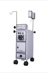
Figure 1: URODYNAMIC STUDY SETUP
The aim of Urodynamic study is to reproduce the LUT symptoms by bladder filling either by infusion catheter or by diuresis as in ambulatory urodynamics. (5)
Uroflowmetry- the patient voids into a flowmeter and flow are plotted with volume on the X axis and time corresponds to Y axis. The uroflowmetry does not reproduce the symptoms as UDS, it suggests voiding dysfunction, simply measures flow. In order to produce results same as with physiological voiding, calm and relaxed environment maintaining patient privacy and dignity need to be maintained. (6)
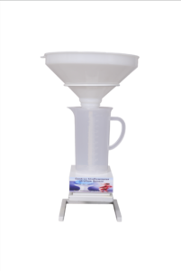
Figure 2: UROFLOWMETRY
Cystometry- is an invasive monitoring by the insertion of catheters into the bladder and rectum. It begins with the near physiological filling rate of bladder. A reduced rate of filling is utilized in bladder pain syndrome and in evaluation of overactive bladder patients. It provides information regarding bladder sensations at various volume as well as the response of the detrusor and urethral sphincter to the bladder filling. It determines the compliance and the capacity of the bladder. The competence of the sphincter on increasing the intraabdominal pressure by coughing, performing Valsalva maneuver and other activity that reportedly causes incontinence in the patient can also be assessed. Abdominal leak point pressure and detrusor leak point pressure are also determined which helps in demonstration of stress urinary incontinence and compliance of bladder respectively. Cystometry ends with a micturition command, or “permission to void”. (7)
A pressure-flow study– the simultaneous measurement of the pressure generated by the detrusor muscle, intravesical and abdominal, volume voided and flow rate. It begins with flow of micturition. It is even more important during the voiding phase to respect the patient’s dignity and privacy and to allow the patient to void in a position that is normal for her specifically sitting. Detrusor activity and urethral function should be defined separately whilst remembering that urethral characteristics will define the precise voiding pressure. Following the pressure-flow study, the post-void residual urinary volume is estimated, and the total cystometric bladder capacity is calculated.(8)
Electromyography- the electric potentials generated by the depolarization of muscle are studied. Potential sampling sites include the intrinsic striated muscle of the urethra, the periurethral striated muscle, bulbocavernosus muscle, external anal sphincter and pubococcygeus muscle. This form of neurophysiological testing may be helpful in diagnosing detrusor sphincter dyssynergia and fowler syndrome. (9)
Urethral pressure profile– valuable diagnostic tool in urinary incontinence,suspected primary sphincter mechanism incontinence, suspected increased urethral tone, reflex dyssynergia, mechanical or functional urethral obstruction and other congenital urethral abnormalities.(10)
Video urodynamics assessment is particularly more informative in neurological patients who have neurogenic bladders and in patients who have had previous surgery or trauma-related anatomical defects. (3)
Ambulatory urodynamics has a portable device continuously monitoring bladder and abdominal pressures via invasive catheters. It is a second line investigation aiming to reproduce symptoms through normal ambulatory activity in patients for whom standard urodynamic testing has not yielded definitive answers. (3)
Standard urodynamic testing involves non-invasive uroflowmetry, followed by invasive cytometry and a pressure-flow study. The addition of tests such as concurrent electromyography (EMG) of pelvic floor muscles and urethral pressure profiles can supplement the investigation for further clinical detail.
References-
- Lenherr, Sara M, and J Quentin Clemens. “Urodynamics: with a focus on appropriate indications.” The Urologic clinics of North America vol. 40,4 (2013): 545-57.
- Bellucci, Carlos H Suzuki et al. “Acute spinal cord injury–do ambulatory patients need urodynamic investigations?.” The Journal of urology vol. 189,4 (2013): 1369-73.
- Clement, Keiran David et al. “Urodynamic studies for management of urinary incontinence in children and adults.” The Cochrane database of systematic reviews vol. 2013,10 CD003195. 29 Oct. 2013
- Nitti, Victor W. “Pressure flow urodynamic studies: the gold standard for diagnosing bladder outlet obstruction.” Reviews in urology vol. 7 Suppl 6,Suppl 6 (2005): S14-21.
- Austin, Paul F et al. “The standardization of terminology of lower urinary tract function in children and adolescents: update report from the Standardization Committee of the International Children’s Continence Society.” The Journal of urology vol. 191,6 (2014): 1863-1865.e13.
- El Helou, Elie et al. “Mobile sonouroflowmetry using voiding sound and volume.” Scientific reports vol. 11,1 11250. 27 May. 2021
- Baines, Georgina et al. “Recent advances in urodynamics in women.” F1000Research vol. 9 F1000 Faculty Rev-606. 15 Jun. 2020
- Drake, Marcus J et al. “Fundamentals of urodynamic practice, based on International Continence Society good urodynamic practices recommendations.” Neurourology and urodynamics vol. 37,S6 (2018): S50-S60.
- Granata, Giuseppe et al. “Electrophysiological study of the bulbocavernosus reflex: normative data.” Functional neurology vol. 28,4 (2013): 293-5.
- Gray, Mikel. “Traces: making sense of urodynamics testing–part 7: Evaluation of bladder filling/storage: Evaluation of urethral sphincter incompetence and stress urinary incontinence.” Urologic nursing vol. 31,5 (2011): 267-77, 289.